Dental · Dental Implants
Subperiosteal Implants in Turkey: A Last Resort for Severe Bone Loss
- Visits
- 2–3 trips (planning scan, surgery with provisional bridge, then the final bridge)
- Days in Turkey
- 2–3 days for scanning if not done at home; 8–12 days for surgery; 6–8 days for the final bridge
- Recovery period
- 2–3 weeks of significant swelling and bruising; soft diet for 8–12 weeks
- Expected lifespan
- Uncertain long term; encouraging short-term data but limited evidence beyond a few years
Want the full picture first?
Read the hub page before comparing single options.
Key facts - A modern subperiosteal implant is a custom 3D-printed titanium frame that rests on the jawbone beneath the gum and carries posts for fixed teeth. - It is reserved for extreme bone atrophy where conventional implants, grafting and zygomatic implants are unsuitable or have failed. - Early studies report high patient satisfaction, but in one multicentre study 65% of patients developed gum recession exposing part of the frame within about 2.5 years. - Indicative pricing is £6,000–£12,000 per arch in Turkey; long-term evidence beyond a few years is limited.
A Last-Resort Option, Explained
Subperiosteal implants in Turkey are increasingly marketed as a "no bone, no problem" solution for patients told they are not candidates for implants. This is one of the most aggressive and least forgiving procedures in implant dentistry, and it should be treated that way.
The concept is old. Subperiosteal implants were developed in the 1940s, when a frame was cast from an impression taken directly on the exposed bone in one operation and fitted in a second. Poor fit, infection and frame exposure made those designs notorious, and they largely disappeared once osseointegrated screw implants became reliable.
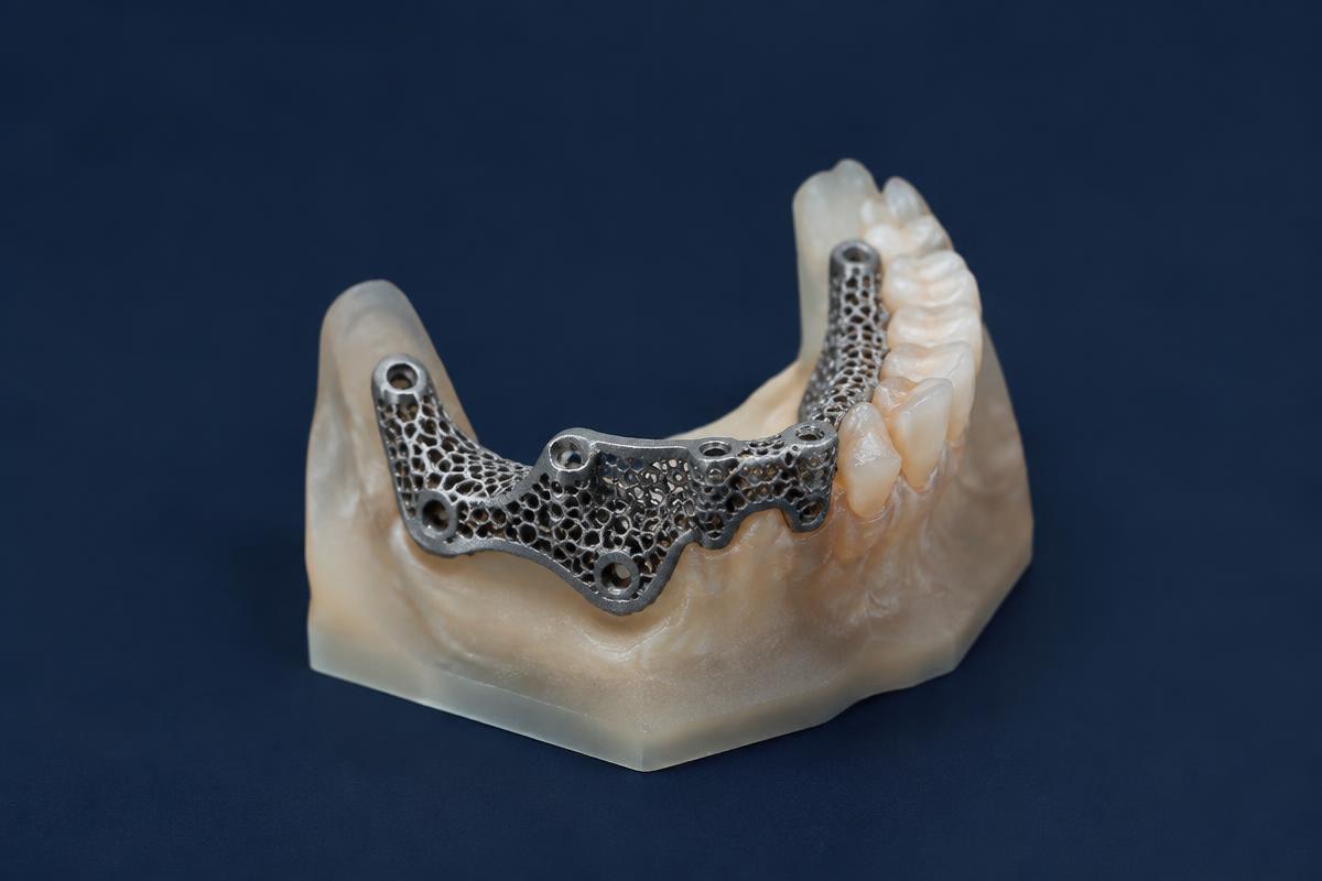
The modern revival, often called the additively manufactured subperiosteal jaw implant (AMSJI), uses CBCT scanning, digital design and 3D-printed titanium. Fit has improved considerably, and early results are encouraging: a prospective one-year study3 and a later 40-patient series reported clear improvements in oral health–related quality of life and high satisfaction.2
The biology, however, has not changed. The frame sits on the bone beneath thin gum tissue, and its success depends on that tissue staying intact. In a multicentre study of 40 patients with upper-jaw frames, 65% developed mucosal recession with exposure of part of the framework after a mean of about 2.5 years. Thin gum tissue and peri-implant inflammation were significant risk factors.1 In a lower-jaw series of 40 frames, two were removed for persistent infection and a third was scheduled for removal.4
Clinically, a subperiosteal implant is indicated only for:
- severe maxillary or mandibular atrophy, where the ridge has resorbed to a pencil-thin or flat form;
- failed grafts or failed zygomatic implants;
- defects after tumour resection.
It is not an alternative for a patient who could have All-on-4, a sinus lift or bone grafting.
Frame Fixation and the Soft Tissue Problem
The typical candidate has severe pencil-thin bone atrophy (Cawood and Howell classes V–VI). In the maxilla, the sinuses may have expanded almost to the ridge; in the mandible, the nerve may lie close to the crest.
The subperiosteal frame does not rely on osseointegration inside the bone. It is fixed with small osteosynthesis screws to the remaining dense cortical bone at load-bearing points:
- in the upper jaw: the piriform rim beside the nose, the zygomatic buttress and sometimes the body of the zygoma and palate;
- in the lower jaw: the chin region and the external oblique ridges, while avoiding the mental nerves.
The lattice surface against bone may allow some bone ingrowth, but the screw fixation provides initial stability. Posts, usually four per arch, rise through the gum to carry the bridge.
The surgery requires wide flap reflection: a crestal incision with vertical releases and a full-thickness flap lifted to expose all the bone the frame will cover. The frame is seated, checked for passive fit and screwed in, then the flap is closed without tension around the posts. This takes about two to four hours per arch under general anaesthesia or deep sedation. A provisional bridge is often fitted within days.
Mucosal dehiscence, the gum breaking down to expose the frame, is the dominant complication. Exposed titanium is colonised by bacteria, which can cause chronic inflammation, infection and sometimes partial or total frame removal. Risk is highest with:
- thin gum tissue;
- flap closure under tension;
- a bulky frame design;
- smoking, poor hygiene or untreated mucositis.
Small exposures can often be managed; progressive ones may need surgical revision.
From CBCT Segmentation to Printed Titanium
Everything begins with CBCT 3D bone segmentation. A high-resolution CBCT, ideally with voxel size of 0.3 mm or smaller and without movement, is imported into planning software, and bone is separated from soft tissue by thresholding. In very thin, low-density bone, segmentation errors creep in: a threshold set wrongly can add or remove fractions of a millimetre, and the frame will not seat.
The segmented jaw is usually 3D-printed, and the frame is designed on it, specifying:
- strut paths that avoid nerves and sinus walls;
- screw positions in the thickest cortical bone;
- post positions dictated by the planned teeth.
The surgeon should review and approve the design before manufacture.
The frame is made as a custom direct metal laser sintered (DMLS) titanium mesh, or by the closely related selective laser melting process, fusing grade 5 or grade 23 titanium alloy powder layer by layer. It is then heat-treated, support structures removed and surfaces finished: rough or porous against bone, highly polished where it passes through the gum. Many workflows also print surgical guides for each fixation screw. Manufacture typically takes two to four weeks.
Planning, Manufacture and Surgery
Stage 1: scan and planning. A high-resolution CBCT is taken, in Turkey or at a verified centre at home, segmented and used to design the frame. The surgeon reviews and signs off the plan and tooth positions.
Stage 2: manufacture. The frame is printed, finished and checked on a printed model of your jaw.
Surgical trip, days 1–2. Pre-operative checks, blood tests and anaesthetic assessment.
Day 3: surgery. The flap is raised, the frame seated and screwed, and the flap closed around the posts.
Days 4–7: provisional bridge. A PMMA provisional is fitted and the bite checked. Swelling is significant.
Days 8–12: review. Suture checks and hygiene training; flying is usually approved by around day 10–12.
Months 3–6: final bridge. The soft tissue around the posts is assessed, scans taken and a titanium-reinforced or zirconia bridge fitted.
Long term. Reviews every six months are essential to catch early exposure, and gum inflammation should be treated promptly, since it is a risk factor for recession.
Red Flags in Subperiosteal Offers
No digital planning or guides. Ask to see the segmented CBCT, the frame design and the printed model, and whether surgical guides will be used for the screws. A clinic that cannot show a surgeon-approved digital plan is not ready for this surgery.
No discussion of alternatives. A clinic that offers only subperiosteal frames will recommend them regardless. A properly assessed patient should hear why zygomatic implants, a sinus lift, grafting or tilted implants are or are not suitable, and what happens if the frame cannot be seated during surgery.
Incomplete pricing and warranty. Indicative costs are £6,000–£12,000 per arch, but headline figures often exclude design and manufacturing fees, general anaesthesia and hospital costs, the provisional and definitive bridges, and management of exposure. Given the reported exposure rates, ask in writing what the warranty covers if the frame is exposed or must be removed.
Candidacy
Ideal candidates
- Cawood and Howell class V–VI atrophy where endosteal implants, grafting and sinus lifts are not viable.
- Failed grafts or zygomatic implants, or defects after resection.
- Non-smokers with good systemic health, thick enough gum tissue and meticulous hygiene.
Contraindications & complications
- Enough bone for conventional implants or All-on-4, including where modest grafting would make them possible.
- Smokers, uncontrolled diabetes, IV bisphosphonates, heavy prior radiotherapy, and thin, fragile mucosa.
- Complications include mucosal recession and frame exposure, infection, screw loosening, nerve disturbance and, in some cases, frame removal.
How It Compares With Zygomatic Implants
Zygomatic implants anchor in the cheekbone, with long-term survival of around 96% at six years in meta-analysis, and allow immediate loading. They are used only in the upper jaw, and risks include sinusitis and, rarely, orbital injury.
Subperiosteal frames can treat either jaw and avoid passing through the sinus, but involve more extensive surgery, thinner long-term evidence and a substantial risk of frame exposure. In the severely atrophic upper jaw, zygomatic implants are generally the better-evidenced first choice. For an explanation of how conventional implants differ, see endosteal implants.
Frequently Asked Questions
I was told I don't have enough bone. Does that mean I need a subperiosteal implant?
Not necessarily. "Not enough bone" means different things for different plans. Many patients turned down for conventional implants can still have tilted All-on-4 implants, short implants, a sinus lift or bone grafting, and in the upper jaw, zygomatic implants have a longer track record. Subperiosteal frames are appropriate only when these options have been assessed on a CBCT and ruled out. Ask for a written explanation of why each alternative is unsuitable in your case, or get an independent second opinion, before agreeing to surgery.
How serious is frame exposure, and can it be fixed?
Exposure is common: in one multicentre study, 65% of patients showed gum recession exposing part of the frame within about two and a half years.1 Small exposures can often be managed with meticulous cleaning, antimicrobial rinses, smoothing the exposed titanium and sometimes a soft tissue graft. Larger or progressive exposures harbour bacteria, cause chronic inflammation and can loosen fixation screws, sometimes requiring partial or complete removal. Thin gum tissue and gum inflammation increase the risk, so six-monthly reviews and prompt treatment of inflammation are essential. Ask how exposure would be managed once you are home.
Why is the CBCT scan so important for this treatment?
The entire frame is designed from the CBCT and cannot be adjusted significantly during surgery. The scan must capture thin, low-density bone accurately; small segmentation errors can prevent the frame seating passively, causing rocking, uneven pressure on the gum and a higher risk of exposure. A good clinic uses a high-resolution scan without movement, verifies the design on a printed model of your jaw, and has the surgeon approve it. If your scan is old, low-resolution or predates further tooth loss, expect a new one.
Can a subperiosteal implant be removed if it fails?
Yes. The frame sits on the bone surface and is held by small screws, so removal is generally less destructive than removing a failed zygomatic implant or a heavily integrated conventional implant. It still requires reopening a flap, removing the screws and lifting the frame, usually under sedation or general anaesthesia. After healing, options usually return to what they were before: a conventional denture, a second custom frame, or zygomatic implants where suitable. Ask whether removal and revision costs are covered by the clinic's warranty.
References
- Van den Borre C, De Neef B, Loomans NAJ, Rinaldi M, Nout E, Bouvry P, Naert I, Van Stralen KJ, Mommaerts MY. Soft tissue response and determination of underlying risk drivers for recession and mucositis after AMSJI implantation in the maxilla. Int J Oral Maxillofac Implants. 2024;39(2):302. PubMed
- Van den Borre C, De Neef B, Loomans NAJ, Rinaldi M, Nout E, Bouvry P, Naert I, Mommaerts MY. Patient satisfaction and impact on oral health after maxillary rehabilitation using a personalized additively manufactured subperiosteal jaw implant (AMSJI). J Pers Med. 2023;13(2):297. doi:10.3390/jpm13020297
- Van den Borre C, Rinaldi M, De Neef B, et al. Patient- and clinician-reported outcomes for the additively manufactured sub-periosteal jaw implant (AMSJI) in the maxilla: a prospective multicentre one-year follow-up study. Int J Oral Maxillofac Surg. 2022. PubMed
- Van den Borre C, Otero JJ, Loomans N, et al. Retrospective analyses of additively manufactured subperiosteal jaw implants in the mandible. Research record